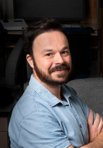
Rafael Arrojo e Drigo, Ph.D.
Assistant Professor
Molecular Physiology and Biophysics Department
Vanderbilt University
Seminar Information

Aging is a biological process characterized by a gradual loss of cell function and increased risk for degenerative and metabolic diseases. While it is generally accepted that most brain neurons are not replaced during an organism’s lifetime, little is known about the longevity of cells in somatic organs. And while the turnover of organelles and protein complexes has been studied at length using bulk mass spectrometry approaches, we lack the understanding on the spatial distribution of protein and organelle longevity in animal models and in situ. Importantly, because of their longevity, long-lived biological structures (e.g., a post-mitotic cell or a long-lived protein) are more likely to accumulate age-related damage that compromises cell function.
During my talk I will describe a multi-modal microscopy approach that combines in vivo stable isotope labelling of mice with correlated electron microscopy (EM) and multi-isotope mass spectrometry (MIMS). This technique, called MIMS-EM, can reveal the spatial pattern of cellular and macromolecular longevity in virtually any tissue or animal model. I will show how we have applied stable isotope labelling and MIMS-EM imaging to map the longevity of nuclear pores, basal body of the primary cilium, mitochondria, hepatocytes, pancreatic cells, neurons, and myocytes. These studies reveal that several types of somatic cells can be as old as neurons and undergo little-to-no turnover during an animal’s lifetime. Likewise, we identify subsets of long-lived mitochondria characterized by the remarkable longevity of proteins in respiratory chain complexes. This pattern of cellular and macromolecular longevity can be vastly heterogeneous, with young and older components sharing the same intracellular and/or tissue microenvironments. We propose a new biological organization pattern called age mosaicism that observed across multiple scales and as a fundamental principle of adult tissue, cell, and protein complex organization. Finally, I will describe recent efforts from our group demonstrating MIMS-EM applications geared towards mapping the spatial organization of nutrient flux in situ to identify novel sub-cellular organization patterns.
Rafael Arrojo e Drigo is originally from Brazil and acquired his Ph.D. at Universidade Federal de Sao Paulo (UNIFESP) in Cellular Endocrinology. Here, Rafael studied under the supervision of Antonio C. Bianco and discovered how the catalytic activation of the hormone T4 by the type 2 deiodinase enzyme (D2) leads to D2 poly-ubiquitination and regulates thyroid hormone signaling and metabolism. For his first postdoctoral experience, Rafael moved to Singapore and joined Per-Olof Berggren’s lab at the Nanyang Technological University (NTU). In Singapore, he developed a high-speed and non-invasive confocal microscopy platform capable of imaging islet blood flow and intracellular beta cell calcium dynamics in mice and non-human primates. In addition, he applied high-resolution light and electron microscopy approaches to identify cellular filopodia adaptations that mediate delta cell somatostatin signaling within the intra-islet environment. Next, Rafael moved to San Diego to join Martin Hetzer’s group at the Salk Institute where he studied beta cell aging patterns. Here, and together with Mark H. Ellisman at UCSD’s National Center for Microscopy and Imaging Research (NCMIR), Rafael developed the multi-scale imaging pipeline called MIMS-EM that combines stable isotope imaging with electron microscopy to quantify cell and macromolecular age in situ. In 2020, Rafael joined the Molecular Physiology and Biophysics department at Vanderbilt University as a tenure-track Assistant Professor. At Vanderbilt, Rafael and his team continue to develop MIMS-EM capabilities and apply a range of imaging and single cell multiomic approaches to investigate the mechanisms regulating post-mitotic cell longevity and nutrient metabolism.
